Lecture number 3 METHODS AND GENERAL PRINCIPLES OF DIAGNOSIS OF TUBERCULOSIS
Clinical symptoms of pulmonary TB are diverse, but the disease has no specific symptoms. This is particularly important in today’s conditions, which are characterized by environmental changes, multiple scattering effects nym on the human body of various vaccines, serums and antibiotics, as well as changes in the properties of the causative agent of tuberculosis.
It should be borne in mind three things:
1) TB patients with symptoms of the disease treated by general practitioners rather than specialist, TB specialist;
2) tuberculosis – an infectious disease, and patients may present to others a serious epidemic threat;
3) treatment of tuberculosis requires the use of specific anti-TB drugs and must be supervised by a specialist phthisiatrician, which owns the necessary knowledge and skills.
Questioning and physical methods of investigation can only be suspected tuberculosis. For early and timely detection of tuberculosis, as well as verified diag Rosa require special methods of investigation.These are the E in Phthisiology are immunological, microbiological, ray, endoscopic and morphological IU ods.They are crucial in the diagnosis and a differential diagnosis of tuberculosis, evaluation of the disease and treatment outcomes.
The main symptoms
Typical symptoms of pulmonary tuberculosis: weakness, fatigue, poor appetite, weight loss, fever, sweating, dry cough or sputum, shortness of breath, chest pain, blood Harkány.These symptoms may be expressed differently and appear in various combinations.
Weakness, fatigue, poor appetite, weight loss, irritability, decreased performance may be early manifestations of TB intoksika tion.TB patients often do not associate these symptoms with the disease, suggesting that their appearance is due through dimensional physical or mental stress.Sim ptomy intoxication require more attention, especially especially in individuals at risk for TB disease.In-depth examination of such patients can be identified by Comrade primary form of tuberculosis.
Increased body temperature (fever) is a typical clinical symptom of infection, and many non-communicable diseases.
In tuberculosis body temperature may be normal, subfebrile and febrile. It is often different pain Scheu lability and increased after physical or psy archical load.TB patients usually suffer fever rather easily, and often almost do not feel.
When tuberculous intoxication in children in the afternoon, the temperature increases for a short time to 37,3-37,5 ° C. These upgrades occur periodically, sometimes no more than J-2 times a week and alternate with long intervals of normal temperature mi.Temperatures rarely pa body remains within 37 ° C, with differences between ut renney and evening temperature is about one degree.Unlike the cases of fever, caused GOVERNMENTAL autonomic disorders, low-grade fever usually declines in tuberculosis in the appointment dopirina s (amidopirinovaya test).Lowering the temperature also occurs against the backdrop of preparations of isonicotinic acid hydrazine.
Steady monotone subfebrilitet low temperature fluctuations during the day is not typical for tuberculosis iron and is more common in chronic nonspecific inflammation in the nasopharynx, paranasal sinuses, biliary tract or genital organs.Body temperature subfebrile can also be caused by endocrine disorders, rheumatic fever, sarcoidosis, lymphogranuloma tozom, kidney cancer.
High fever hectic type is highly characteristic of acute progressive tuberculosis and severe lesions (miliary tuberculosis, caseous pneumonia, pleural em Pius).Intermittirutoschaya hectic fever is one of the diagnostic features that distinguish the form of miliary tuberculosis typhoid from typhus. Unlike tuberculosis, of typhoid fe Tee’s body temperature has steadily increased and then maintained consistently high for a long time.
In rare cases, patients with pulmonary tuberculosis note perverted type of fever, when the morning temperature is above you night.Such evidence of severe fever Institute intoxication, which may have a non-tubercular origin.
Increased sweating is a very common sign of intoxication. TB patients at the beginning of the disease is often noted increased sweating of the head and chest at night or early morning hours. Pronounced sweating (a symptom of a “wet pads”) in the form of profuse perspiration happens in caseous pneumonia, miliary tuberculosis and other severe and its complicated forms, as well as nespetsifi České acute infectious diseases and acute exacerbation of chronic inflammatory processes.
Cough is often accompanied by inflammation, cancer and other lung diseases, respiratory tract, pleura, mediastinum.
In the early stages of TB can cough Jet absent.Sometimes the patient noted a small, ne periodically occurring cough.With the progression of tuberculosis cough increased. It is a dry (not productive) or sputum (productive).Dry paroxysmal cough occurs when compression of a bronchus or enlarged lymph nodes removed GOVERNMENTAL bodies mediastinum.Such a shift is possible if large quantities of fluid in the pleural cavity in patients Nogo exudative pleurisy.Particularly common in dry stupoobrazny cough occurs in tuberculosis bronchus.A productive cough appears in patients with pulmonary tuberculosis in cases of destruction of lung tissue, education nodulobronhialnogo fistula, rupture into the bronchial tree of fluid or pus from the pleural cavity. TB cough leze also may be due to nonspecific chronic bronchitis or bronchiectasis.
Sputum of patients with an initial stage of tuberculosis is often non-cross linked allocation associated with chronic bronchitis ical.After the collapse of a lung Noah fabric of sputum increases.When neoslozh nennom pulmonary tuberculosis sputum is usually colorless, homo gene and has no smell.Accession of non-specific inflammation leads to increased cough and a significant increase crease in sputum.In these cases, purulent sputum may become the character.
Dyspnea is a clinical symptom of respiratory or cardiovascular failure. When the disease is more likely s lungs caused by a decrease in the respiratory surface, a violation of bronchial permeability, limited excursions of the chest wall and diaphragm, a violation Niemi transport of gases through the wall of the alveoli.Certain important effects on the respiratory center GOVERNMENTAL toxic products generated during the life of pathogens and the decay of tissues.
Severe apnea is usually observed during acute pulmonary tuberculosis, as well as chronic disseminated, fibrocavernous, cirrhotic pulmonary tuberculosis.
Progression of tuberculosis and other lung diseases can lead to chronic pulmonary CERD sample and cardiopulmonary diseases.In these cases, dyspnea markedly increased.
Shortness of breath is often the first and main symptom of pulmonary complications such as spontaneous pneu motoraks, atelectasis, or the proportion of lung embolism in the pulmonary artery.With a significant and rapid accumulation of fluid in the pleural cavity in shortness of breath can occur suddenly and be pronounced.
Chest pain is a symptom of the trachea, lung, pleura, heart, aorta, pericardium, chest wall, spine, esophagus and sometimes the abdominal cavity.
Pulmonary tuberculosis chest pain usually caused by spread of inflammation in pa rietalnuyu pleura and the development of perifocal adhesive pleurisy.The pain occurs and increases in breathing, coughing, abrupt movements. Localization of pain usually corresponds to the projection of the affected part of the lung to the chest wall. However one in inflammation diaphragmatic and mediastinal pleura pain radiating to the epigastrium, neck, shoulder, heart area.
In the dry tuberculous pleurisy pain occurs ispod wills and stored for a long time.It increases with coughing and deep breathing, with pressure on the chest wall, and depending on the location of inflammation may radiate to the epigastric or lumbar region, which leads to diagnostic difficulties. In patients with exudative pleurisy tuberculosis chest pain occurs acutely, but with the accumulation of pleural fluid decreased and remained dull until it resorption.
Tuberculosis and other inflammatory lung diseases characterized by weakening and disappearance of pain, even without recourse of the underlying disease.
In cases of acute pericarditis, which occurs in tuberculosis, often a dull pain, unstable. It reduces Xia patient in a sitting position with tilt forward.When a pericardial effusion in the pain subsides, but its disappearance may occur again.
Sudden sharp chest pain occurs when the complication of spontaneous pneumothorax tuberculosis. In contrast to the degree Knockard and myocardial infarction pain in pneumothorax increases during the talk and cough, not radiating to left arm.
If intercostal neuralgia pain is limited by the localization tion course intercostal nerve and increases with the pressure on the intercostal area of the gap.In contrast to the pain of pleural tuberculosis patients, it also amplified Vaeth at an inclination of the body in the affected side.
Lung cancer and other tumors of the chest pain continued and may gradually increase.
Hemoptysis or pulmonary hemorrhage is more common in the infiltrative, fibrous-cavernous pulmonary tuberculosis and cirrhotic.Usually it is phased out etsya, and after the separation of fresh blood for several days continued expectoration of dark clots.In the case of postgraduate radio and blood of aspiration pneumonia after hemoptysis may increase body temperature.
Hemoptysis is also observed in chronic bronze hit, and many nonspecific inflammatory, neoplastic and other diseases of the chest.Unlike tuberculosis, in patients with pneumonia usually occurs first chills, increased body temperature, and then there hemoptysis and stabbing pain in the chest. At Institute farkte lung initially appears more chest pain, then fever, hemoptysis occurs.The duration of Noah hemoptysis seen in patients with lung cancer.
Massive pulmonary hemorrhage more often in patients with fibro-cavernous, cirrhotic tuberculosis and gangrene of the lungs.
In general, one should bear in mind that pulmonary tuberculosis often starts as a common infectious disease with symptoms of intoxication and often takes place under the weight of Koi influenza or pneumonia.The treatment of broad-spectrum antibiotics, the patient’s condition may improve sewing.Further course of tuberculosis in these patients usually wavy: periods of exacerbation displacement nyayutsya remission and periods of relative prosperity.When extrapulmonary forms of tuberculosis, along with general symmetry ptomami resulting from tuberculous intoxication in patients with marked and local manifestations of the disease: in tuberculous meningitis – headache, laryngeal tuberculosis – a sore throat and hoarseness, with osteo-articular tuberculosis – back pain or joint, and changes in stiffness of gait, with tuberculosis of female genital organs – pelvic pain, menstrual function, with tuberculosis of kidneys, ureters and bladder pain in the lumbar region, dizuricheskie disorders, tuberculosis mesenteric lymph nodes and intestinal abdominal pain and disorders function as ludochno tract.Often, however, patients with extrapulmonary tuberculosis, especially in the initial stage, not predyav lyayut no complaints, and disease detected only by special methods of investigation.
Questions, methods fizikalnys
A history to determine when and how you have been revealed by the disease: when you contact a doctor about any complaints or follow-up examination.The patient was questioned about the time of onset of symptoms and their dynamics namics, previously an illness, injury, operations.Pay attention to possible manifestations of tuberculosis such kuleza as pleurisy, lymphadenitis.Detect the disease at which the risk of tuberculosis is particularly high: diabetes mellitus, silicosis, stomach ulcer and duodenal ulcer, alcoholism, drug addiction, HIV infection.
Important information about staying in areas with particularly high rates of tuberculosis, the participation in hostilities, the patient stays in the city or rural areas NOSTA.Are important data about the profession and the nature of the paper, the material conditions of life, lifestyle, alcohol consumption, smoking, as well as stay in the facility s prison system.Assess the level of culture the patient. Parents of affected children and adolescents rasspra shiva on anti-vaccination and the results that berkulinovyh samples.It is also necessary to obtain information tion on the health of family members, possible contact with TB patients at home, attending college, at work and its duration, the presence of TB animals.
Examination.Not only medical but also in the literature describes the appearance of patients with advanced pulmonary tuberculosis schim, which is known as habitus phftsicus.This is underweight, blush on his pale face, bright eyes and wide eyes, dystrophic skin changes, long and narrow thorax, dilated intercostal spaces, acute epigastric angle, lagging (krylovid nye) blade.These external signs are usually seen at far come of tuberculosis. On examination, patients with incipient tuberculosis of any pathological changes sometimes do not show. However, inspection is always required. He often reveals a variety of important symptoms and should be held to the floor constant volume.
Pay attention to the physical development of the patient, the color of the skin and mucous membranes. Compare the severity of the subclavian and pits, symmetrical right and left halves of the chest, assess their mobility during deep breathing, breath support participation in the muscles. Note the narrowing of the intercostal or extension of the interval, post-surgical scars, fistulas, or scars on their healing follows.On the fingers and toes paying attention to the deformation of the terminal phalanges in the form of drum sticks and changing the shape of the nails (in the form of convex time degree count).In children, adolescents and young adults visiting the Ute on the shoulder of scars after BCG vaccination.
Palpation to determine the degree of humidity or dryness of the skin its turgor, the severity of the subcutaneous fat layer.Carefully palpates lymph nodes in the neck, armpits and groin areas. In the inflammatory processes in the lung involving the pleura are often lagging behind the affected part of the chest during breathing research institutes, breast soreness.Patients with chronic tuberculosis and after major operations may be atrophy of muscles of the shoulder girdle and chest. Noe significant mediastinal shift can be determined by palpation on the Status of the trachea.
Voice tremor in patients with pulmonary tuberculosis is common, reinforced or weakened. It is better conductivity converges on the areas packed with infiltrative lung and cirrhotic tuberculosis of the large cavity with a wide draining bronchus.Weakening voice shaking until his disappearance in the pres ence observed in the pleural cavity of air or fluid, atelectasis, pneumonia with massive bronchial obturation.
Percussion reveals relatively coarse changes in the lungs and chest with infiltrative lesions of cirrhotic or equity nature fibrosis membrane ry.The important role played percussion in the diagnosis of non-negative spurious states, as a spontaneous pneumothorax, severe pleural effusion, atelectasis of the lung.The presence of Nogo koroboch or shortened pulmonary sound allows you to quickly assess the clinical situation and conduct the necessary studies.
Auscultation. Some respiratory diseases, especially tuberculosis, may not be accompanied by a change in the nature of respiration and the appearance of additional noise in the lungs. One reason for this is obturation bronze Hove, draining the affected area, dense caseous necrotic masses.
The weakening of breathing characteristic of pleurisy plevral GOVERNMENTAL adhesions, pneumothorax.Rigid or bronchial breathing can listen over infiltrated le gochnoy cloth amforicheskoe – the giant cavern with a wide draining bronchus.
Rales in the lungs and pleural friction rub is often help you diagnose this pathology, which is not always detected by X-ray and endoscopic studies research institute.Finely rales on a limited teaching are a sign of dominance stke effusion of the component in the area of inflammation, and medium and large bubbling rale – a sign of decay or cavities of the cavity.For vyslu Shivani rales should ask the patient to cough after deep breaths, a short pause, and then again a deep breath.At the same time at the height of a deep breath or wheezing occur increases their number. Dry rales are bronchitis, whistling in the bronchi those with bronchospasm.In dry pleurisy auscultated pleural friction rub, and pericarditis – pericardial friction noise.
Microbiological studies
Microbiological and, in particular, bacteriological studies are needed to:
- identify the most dangerous in terms of an epidemic of tuberculosis patients;
- verification of the diagnosis of tuberculosis;
- determine the treatment strategy (organizational form, treatment plan);
- assess the effectiveness of treatment and prognosis;
- for epidemic control of tuberculosis. The objects of bacteriological examination are various fluids and tissues obtained during the examination of respiratory organs.The most frequently examined sputum. Other materials are mi respiratory secretions after aerosol inhalation, bronchoalveolar washings, pleural fluid, gastric washings (mainly in children who do not expectorate sputum and zaglaty vayut).During and after surgery are taken material for the study of rezektaty lungs, pleura, lymph nodes.
In patients with extrapulmonary TB or suspected such explore the various fluid – cerebrospinal, pericardial, synovial, ascitic, and menstrual blood, pus, bone marrow punctate, granulation, scrapes synovial membranes, lymph nodes and punctate, rezektaty bodies.
Classical methods of bacteriological examination are smear microscopy diagnostic material and culture (culture) with the identification of species of microorganisms.
In identifying the IMT should determine their sensitivity to chemotherapeutic drugs.
Smear microscopy diagnostic material – a mandatory method of research in medical uch rezhdeniyah obshey health care services.The purpose of the study – showed tion of acid-fast bacilli.The method we conclude etsya in direct light microscopy of smears stained with Ziehl-Nelsenu, or fluorescent microscopy with approx Rusk flyuorohromnymi dyes.With tuberculosis went FIR is primary, tentative study identifies those patients who are most dangerous in regard to epidemic.
If you find questionable or acid-fast bacilli by microscopic examination of the primary patient sent to the institution for verification of TB diagnosis.
In the bacteriological laboratories of TB services in sputum negative results of the study, not once, but for 3 consecutive days or more. For the study of collecting morning portion of sputum.
Previously the patient should brush your teeth and rinse your mouth. In the sealed vial to collect 3-5 ml of sputum, but the number is valid and less Excellency.In the absence of wet you may use aerosol irritant inhalation of 5-15% solution of sodium chloride dissolved in 1% solution of sodium bicarbonate.Mate rials for research in such cases may be
Sputum collection for the prevention of airborne infection is produced in a dedicated room.
When stained by Ziehl-acid-fast bacilli Nelsenu painted in red, and the surrounding background and acid-fast bacteria in blue. The resolution of the microscopic method revealed acid-fast bacilli leniya increases significantly with fluorescent microscopy with fluorochrome stain pre Parata – auramine or auramine with rhodamine.When illuminated with ultraviolet light mycobacteria tyatsya bright yellow on dark green background.
However, the microscopic method has its limits. Straight (Easy) microscopy with stain Ziehl-Nielsen well can detect mycobacteria in their content of more than 5000-10000 microbes in I ml of the material.Such a number of bacilli found in sputum of patients with advanced tuberculosis Thus.With fluorescent microscopy can reveal the MBT in their content of about 1,000 per 1 ml.
In the early stage of Mycobacterium below detection capabilities Skim microscopic study.In addition, microscopy enables us to differentiate not Mycobacterium tuberculosis with other mycobacteria (mycobacteriosis agents) and, therefore, is insufficient to reliably determine the etiology of the disease.
Disadvantages of the method fills the microscopic Skogen culture methods, or method of planting material for culture media.To obtain a culture of this method is sufficiently MW from 20 to 100 bacterial cells in 1 ml of the ILO, the liquid material.
There are also semi-liquid and liquid nutrient media. Some of the methods with the use of highly selective media can cultivate a culture of mycobacteria in a shorter time, but to identify the organism need more time.
Obtaining a pure culture of mycobacteria by biochemical methods can distinguish M. tuberculosis from other non-tuberculosis mycobacteria, to determine their viability, virulence and drug susceptibility nym drugs.It is also possible to quantify bacterial surround: lean-to 10 colonies on the medium, mild Noah – from 10 to 50, and abundant – more than 50 colonies.
The disadvantage is its culture-length – about 2 months. However, this method should be used in all patients with suspected tuberculosis, especially particularly with repeated negative results of microscopic examination.
In order to expedite the study were aware of the culture apparatus for the radiometric determination of the growth of E kobaktery.In devices of this type of liquid nutrient medium but contains 14 C-labeled palmitic acid.Of a special sensor detects a sign of growth of mycobacteria in the allocation of 14 C within 10-12 days or sooner.
Another principle is based on the absorption of oxygen in the growth process of the microbial population. Reducing the concentration of oxygen in a closed chamber activates a fluorescent indicator which glows under ultraviolet light.
By using such devices as possible identification of mycobacteria and the determination of their sensitivity to medicinal preparations vennym.Developed and committed correspond to other automatic devices.
Cultural studies to determine the sensitivity of the Office to the drugs and to identify resistant strains of them.
Considered sensitive mycobacteria, which ceased to grow and multiply when you add in the nutritional environment of the drug in relatively low, ie, the critical concentration.At the high stability of mycobacteria in the drug concentration that does not work.
A quick way to identify resistance to drugs MW can be considered the use of biological microchips.They are used to estimate the kinetic re DNA polymorphism.
Small size, high sensitivity and specificity of biological microchips allow you to apply them simultaneously to identify the causative agent and determine its resistance to several drugs nym drugs.The result of such investigations have generally followed the traditional culture-control method.
Laboratory determination of the spectrum and the degree of resistance to Mycobacterium tuberculosis drugs is of great importance for the choice of chemotherapy, monitoring the effectiveness of treatment and epidemiological monitoring.
Bacteriological examination in low information content can be supplemented by biological methods home.It is the infection of an animal obtained from the pain Nogo biological material, which presumably contains but the ILO. Usually used in guinea pigs.These animals are highly sensitive to the Office.
About a month after infection in pigs, generalized ny tuberculosis.The biological method is vysokochuvst Indeed, as tuberculosis in the guinea pig occurs with the introduction of a material containing at least 5 of bacteria in 1 ml.However, it is complex in terms of organization and requires a fairly high cost.
In difficult diagnostic cases, resorted to molecular methods give biologicheskkm study.They are insulated amplifika function, the system is insulated amplification of sequences of the target, ligase chain reaction, polymerase chain reaction (PCR).
The most widely used PCR with primers specific for IMT. The reaction is based on amplification of specific DNA M. tuberculosis.PCR – a highly sensitive and rapid method for laboratory diagnosis of tuberculosis iron.Identification of the Office of the diagnostic material in the presence of 10.1 cells in a sample can be carried out for 5 – 6 pm For PCR requires special test systems and laboratory topics.
The study of blood and urine
In patients with pulmonary tuberculosis changes in the overall analysis of blood se are not pathognomonic.In the acute stage of the disease observed low leukocytosis, lymphopenia, monocytosis, left shift leukocyte (a significant increase in percentage of stab neutrophils), increased ESR crease. In patients with advanced tuberculous lesions and severe intoxication, sometimes a hypochromic anemia.The involution of tuberculous inflammation leniya celebrate the normalization of white blood cells, lymphocyte count reaches the norm and even increased.
Biochemical blood analysis produced estimates for the phase and the characteristics of a tubercular inflammation of the liver functional definition and exclusion of associated diabetes mellitus.
In acute tuberculous inflammation diminishes the albumin-globulin coefficients, increases in plasma fibrinogen and sialic acid, there is C-reactive protein.In patients with a large number of sputum, copious purulent exudate General of membrane, renal amyloidosis can be observed hypoproteinemia.
Determination of blood content of alanine and aspartic aminotransferase, alkaline phosphatase, bilirubin, residual nitrogen, urea, creatinine, and coagulation tests (thymol, sulemovaya) give evidence of liver function and tolerability of drug therapy. In order to prevent tuberculosis is often associated with diabetes self Harney to determine blood glucose and glycated hemoglobin.
Immunological blood test sometimes used to confirm the tuberculous etiology of the disease, its activity of Definition and evaluation of the effectiveness of treatment.Used for this ELISA, which oc Nova antigen-antibody reaction.Antibodies to the IMT found lyayut with a special preparation of tuberculin – a diagnosticum erythrocyte antigenic dry.Sequence sensitive ELISA in tuberculosis is 60-70% and specificity – 90%.
With active tuberculosis often exhibit positive reactions blast transformation of lymphocytes and inhibition of leukocyte migration in terms of their stimulation of the Kulin tuberculosis, as well as reducing the number of immune cells.The results of these immunological studies of depend on the phase of tuberculosis.In patients with progressive siruyuschim course of TB is most pronounced inhibition test migration of leukocytes.An exacerbation of inflammation spec REFLECTION reduced number of T-and increases the number of rosette-forming lymphocytes B with a simultaneous decrease in the content of G – and A-immunoglobulins.For involution of tuberculosis characterized by severe blast transformation reaction of lymphocytes in the presence of tuberculin PPD.
Immunological tests used for differential diagnosis of tuberculosis.For example, in cancer and sarcoidosis, in contrast to tuberculosis noted suppression of T-lymphocyte blast transformation in response to fitogemagtlyu-tininom.
The overall clinical analysis of urine from patients with tuberculosis occasionally reveals a serious complication of major diseases.Thus, in severe tuberculous intoxication in the model Th may appear white and hyaline cylinders.Complications tion of pulmonary tuberculosis with amyloidosis leads to persistent proteinuria and microhematuria.Of tuberculosis in kidney can detect human protein, white blood cells, red blood cells are often, but some patients also MW.
Tuberculin
Reaction to the introduction of tuberculin used as a diagnostic test to detect specific sensitization of the human body to the IMT. It is called tuberculin, and is widely used in mass screening for tuberculosis and for individual diagnosis in clinical practice.
Mass tuberculin survey called the large groups of people, usually children and adolescents, through tuberculin tests for epidemiological, clinical and diagnostic data.
Individual tuberculin used for individuals in specific clinical indications.
Tuberculins. Tuberculin was obtained by Koch in Berlin in 1890It was a special water-GRAIN tannuyu glycerin extract of the culture of the IMT and later was named the old, or alttuberkulinom (German – Alttuberculinum Koch).Old tuberculin Koch included a lot of ballast substances from the culture medium on which the cultured mycobacteria. With the presence of these substances, mainly proteins, bind the frequent occurrence of nonspecific reactions GOVERNMENTAL time with the introduction of old tuberculin.
In 1934F, Seibert, and S. Glenn of Philadelphia created a pure drug-Purified protein derivative (PPD-S).In our country dry purified tuberculin was manufactured in 1939 at the Leningrad Institute of Vaccines and Sera, under the guidance of MA Linnikovoy and became known as PPD-A.
On the biochemical composition of tuberculin is a complex mix of tuberkuloproteinov, polysaccharide, lipid fractions, nucleic acids. It contains the remains of bacteria and waste products of the causative agent of tuberculosis.
With immunological products tuberculin is specific allergen and incomplete antigen containing individual antigenic components of the IMT.
The drug is dosed in conventional tuberculin units – TE. In accordance with the international standard for a TE accept such amount of tuberculin to which the positive-reacting 80-90% of infected people.In a TE 0.00006 mg dry preparation PPD-L or 0.00002 mg PPD-S.
In Uzbekistan, produced two types of tuberculin PPD-L:
– Purified tuberculin in standard breeding – a vial with 3-5 ml in 0.1 ml of which contains the PPD-A with 2m;
– Dry purified tuberculin (ALT) in ampoules of 10 000 TE, which is to be breeding in the attached carbonated isotonic sodium chloride solution.
Purified tuberculin in standard dilution of PPD-A with 2m of 0.1 ml is used for mass and individual tuberculin skin test in the test.
Dry purified tuberculin used only in specialized TB facilities for different tuberculin tests.
Methods of tuberculin tests. There are three methods of administration of tuberculin – skin, intradermal and podkozh ny.They are used for subcutaneous samples Koch (1890) dermal test Pirquet (1907), intracutaneous test Mantoux (1909).
A sample of Koch with subcutaneous administration of tuberculin used in TB hospitals in the differential diagnosis of tuberculosis and determine the degree of activity that berkuleznogo process.For the selection of pre-determined dose of tuberculin sensitivity threshold to him by titration with sequential increase in dose. Tu berkulin injected subcutaneously in the upper third of the shoulder, sometimes in the angle of the scapula.The dose of tuberculin in children is usually 10-20 TE, TE in adults -20-50. The sample Koch should be made with caution, since it can provoke the progression of tuberculosis.
Sample Pirquet currently has very limited application of Noah.Usually used as a modification of its cutaneous tuberculin test graded.
Used a sample of hospitals in TB patients with tuberculosis of children and adolescents to determine the indie vidualnoy tuberculin sensitivity.To perform epicutaneous graded sample solutions are used for TB Culina at different concentrations: 100, 25, 5 and 1%.The sample was produced on the skin of the forearm. Tuberculin applied to the skin drops, and then through the skin Katsila scarifying.
The most common is the intradermal Mantoux test. It is made by a physician specially trained health Skye nurse or paramedic with document-tolerance for the tuberkulinodnapyustiki.Mantoux test usually performed on an outpatient basis.
For the Mantoux test using special tuberculin syringes high disposable 1 ml and fine short needle with a sharp oblique cut.In gaining a syringe 0.2 ml of tuberculin and released from a 0.1 ml, so that the volume of drug injected was 0.1 ml (2m). The inner surface of the middle third of the forearm skin area treated with 70 ° ethanol and dried. The needle is introduced into a cut up the surface layer of stretched skin (intradermally) in parallel to its surface. After the dive holes ticipation needle into the skin from a syringe injected 0.1 ml of TB lin.With proper technique in the skin a papule is formed in the form of lemon peel de whitish with a diameter of at least 7 – 9 mm.
In the mass screening for tuberculosis intradermal tuberculin used only the Mantoux test with 2 tuberculin units of purified (PPD-A with 2 TE) in the standard dilution.
The reaction to the introduction of tuberculin. Office Infected persons with tuberculosis or have had it before, as well as in BCG-vaccinated, in response to the tuberculin allergic reactions caused by PCHZT. This reaction is highly specific and occurs only in response to the introduction of the antigen, which was September sibilizirovan the human body.PCHZT reaction starts to form after 6-9 h and reaches its maximum expression zhennosti 72 hours after injection of tuberculin.
The severity of local, focal and general reactions to the tuberculin dose-dependent and the method of administration, degree of sensitization of the organism, its general reactivity.
Local reaction to the introduction of tuberculin in sensitized Office organism occurs when skin, intra-dermal and subcutaneous injection of tuberculin.It usually shows up at the site of tuberculin erythema and form valuations of infiltration – papules.Take me sometimes formed molecule, ulceration, there are signs of local limfan Gita and regional lymphadenitis.Focal reaction in the form of acute inflammation of peripheral focal observed in patients TB patients with subcutaneous administration of tuberculin.On schaya or systemic reaction manifested deterioration, fever, changes in hemogram and other recognition kami amplification of tuberculosis intoxication.The overall reaction occurs in patients with tuberculosis in the subcutaneous tuberculin and in some cases its intradermal administration.
Evaluation of the results. The sample Koch estimated 48-72 hours and is considered positive if there are signs of focal and general response of an organism. The general reaction ha acterized by malaise, fever, changes in hemogram parameters and protein composition of blood.Focal reaction in pulmonary tuberculosis expressed in the appearance or exacerbation of wheeze, increased infiltration around lesions possible discovery by the Office in the sputum. Kidney observed in tuberculosis Pyuria and the Office of the urine, tuberculosis eye – an increase in area of inflammation. The local reaction in the test Koch’s tuberculosis patients usually shows infiltration with 10 – 20 mm.Compared in comparison with the general and focal reactions it has less diagnostic value.
The sample Pirquet evaluated after 48 h and considered positive with a diameter of 3 mm and infiltrate more than 100% for TB Kulin.A positive response to all concentrations of TB usually ling evidence of active primary tuberculosis.
The result of the Mantoux test evaluated after 72 hours of a doctor, nurse or timeshare Feld, who carried out the sample.For this purpose a transparent ruler with millimeter divisions are measured and recorded transverse to the axis of the forearm the size of the infiltration tus.In the absence of infiltration is measured and recorded congestion.
Reaction to the tuberculin skin test when the sample is evaluated by the following criteria:
- Negative – infiltrate and hyperemia absent;
- Questionable – infiltrate 2-4 mm in diameter, or hyperemia of any size;
- positive – infiltrate a diameter of 5 mm or more.If you infiltrate a diameter of 5 – 9 mm reaction considered weakly positive, 10-14 mm – medium intensity, 15-16 mm – marked.
In children and adolescents in a reaction called hyperergic infiltrate a diameter of 17 mm or more, and in adults – 21 mm or more.Other signs hyperergic reactions may be the appearance of vesicles, ulceration, or regional lymphadenitis lymphangites – regardless of the amount of filtrate Institute.
Under mandatory for children and teenagers in the skin, vaccination and revaccination with BCG a positive Mantoux test with PPD-A 2m can testify as an infectious and postvaccinal of allergies.
To distinguish between them account for the intensity of tuberculin reaction time from the last BCG vaccination, the presence and size of post-vaccination scar, as well as the possibility ny contact with TB patients and the presence of clinical symptoms.For postvaccinal allergies ha characteristically suspicious and expressed mild reaction with a diameter of 2.11 mm infiltrate.More pronounced reaction to the tuberculin skin infiltrate with a diameter of 12 – 16 mm are in re-vaccinated children and adolescents, in the presence of large post-vaccination scars (6-9 mm or more).Post vaccination allergy when re-sample at 3 months tended to weaken.
Children with frequent clinical manifestations of allergy nespetsifi Ceska Mantoux test is recommended after the desensitizing treatment.Saving tuberculin sensitivity at the same level or increased during treatment confirm the infectious nature of the allergy.
A negative Mantoux test indicates anergy, which can be positive or negative.
Positive anergy observed in uninfected Office of healthy subjects and negative in patients with progressive course of tuberculosis, infected at the Office of HIV infection and with different comorbid conditions.
Dynamic assessment of tuberculin sensitivity by the Mantoux test result makes it relatively quickly identify the time of primary infection with the IMT. To this end, Russia tuberculin sensitivity in children and young shoots examined a once a year.The results of the corresponding record in the individual section maps of ambulatory monitoring.
The appearance of the year the first positive reaction to the tuberculin skin test in the test ATD-A with 2 TE in unvaccinated child or young person is called superelevation tuberculin sensitivity.
In the context of widespread vaccination of infants and tuberculosis revaccination of children and adolescents in terms decreed a sign of a bend sensitivity to tuberculin is considered to be strengthening the response to tuberculin reac tion at regular Mantoux test.This gain manifests increasing diameter of infiltrate at the site of the Introduction to the PPD-A 2m by 6 mm or more compared to its size a year ago, as well as signs of giperergii to tuberculin.
Virage tuberculin sensitivity confirms the initial infection took place MBT and is the basis for the complex medical and preventive České events.They are aimed at preventing the progression of primary infection with the development of symptomatic tuberculosis and the detection of sources of infection.
According to the results of the Mantoux test positive IMT include children and adolescents with the following features:
- an annual observation was first noted by a positive reaction (papule diameter of 5 mm or more), not associated with BCG vaccination;
- Within 4-5 years, steadfastly maintained a positive reaction with infiltration of 12 mm or more;
- In the year since the tuberculin sensitivity increased dramatically – the diameter of the infiltrate increased by 6 mm or more;
- Within a few years there has been increased tuberculin sensitivity with increasing diameter of the infiltrate to 12 mm or more.
Virage reaction to tuberculin, long-term preservation of a positive reaction in the presence of infiltrate a diameter of 12 mm or more, hyperergic reaction and increasing the reaction is the basis for the inclusion of healthy children, adolescents and adults in a group of individuals at increased risk of tuberculosis.
In assessing the Mantoux test results should take into account the presence of concomitant infectious diseases (measles, chickenpox, whooping cough) or somatic pathology (capkoidoz, asthma, rheumatism, malignant but voobrazovaniya).The response to tuberculin reaction function is also dependent on the allergic disposition of the body, the phases of the ovarian cycle in women., The individual sensitivity of the skin.We can not exclude the role of exposure to environmental hazards in the form of high background radiation, emissions of chemical plants, etc. At the end, the result of tuberculin skin test may be affected by irregularities in her conduct, as well as incorrect storage of tuberculin.
Indications and contraindications.Mass tuberculin used for early detection of the primary Institute fitsirovaniya MW and TB, the definition of indications for revaccination, epidemiological surveillance of tuberculosis.To all of the BCG-vaccinated children (from 12 months of age) and teenagers hold annual Mantoux test with 2 TE – regardless of its previous of the result.Infants who are not vaccinated with BCG vaccine in the neonatal period for medical contraindications, the Mantoux test done 6 months of age, 2 times a year (before instilling ki BCG-M).Conduct tuberculin skin test beam above in one season, mostly in the fall.When suspicious rhenium in the primary infection of children with superelevation reaction to tuberculin, with hyperergic or increasing sensitivity to tuberculin should be sent to the TB specialist.
Indications for BCG revaccination identify children at age 7 (grade 1) and adolescents in the 14 loop (7-8th grade). In healthy subjects with a negative reaction to the tuberculin (Mantoux test with 2 TE) consider re shown the vaccine.
On the epidemiological significance of mass tuberculin position is to determine the percent of infected people in large groups of the population and estimates of the annual risk of infection with the IMT. This figure reflects the percentage of newly infected individuals for use streamed year.This includes children and adolescents with superelevation tuberculin sensitivity and persistent high or increasing reaction to tuberculin.
Mantoux test with PPD-A 2m harmless to healthy children, adolescents and persons with various somatic diseases niyami.However, some previous diseases and immunizations may strengthen or weaken the skin sensitivity to tuberculin children. In this regard, skin diseases, infectious onnye, an infectious-allergic and somatic diseases
b exacerbation, allergic conditions are about tivopokazaniyami to the Mantoux test in mass tuberculin.The sample was not done in 1 month after any preventive vaccine or biological diagnostic tests skoy, as well as quarantine for childhood infections.
Mass tuberculin conduct medical vention about general health care facilities.Methodological guide skoe mass tuberculin impl stvlyayut TB dispensaries.If done right at an annual tuberculin diagnosis should be changed in 90-95% of child and adolescent population.
In children’s groups organized a mass tuberculin diagnosis by trained medical performs sky staff, other children and young preschoolers Mantoux test carried out in a children’s clinic.In rural areas, carry out tuberculin district and district health centers, and in some cases – midwife posts.
Individual tuberculin diagnosis by means of the Mantoux test with 2 TE is used for the differential diagnosis of infectious diseases and allergy to tuberculin postvaccinal to monitor the status of persons at increased risk of tuberculosis.Other indications explosion lyayutsya diagnostic problems in cases of many diseases, which should be differentiated from tuberculosis,
In the tuberculin tuberculosis institutions use to determine the activity of tuberculous process, assess the effectiveness of treatment and diagnosis of tuberculosis locally.Indications for such probes is determined on the basis of clinical data, regardless of the previous limitations of the sample. Mantoux test is conducted not only with 2 TE, but also with other doses of tuberculin. Also use a graded test Pirquet test and Koch, sometimes defined lyayut tuberculin titer produce eosinophil-tuberculin or tuberculin skin test-gemobelkovo.When these tests assess the body’s response to tuberculin of the corresponding changes in blood tests.
Contraindications for tuberculin, but are hypersensitive to tuberculin, no.
Assessment of circulatory and respiratory functions
Pulmonary tuberculosis, particularly chronic and widespread, usually accompanied by impaired respiratory function and changes in respect to the cardiovascular system.They may be due to intoxication, damage to eat lungs, pleura, bronchi.
Investigation of circulatory and respiratory functions are usually not important to establish the nosological diagnosis, but it is undeniable role in assessing the overall state of the patient, the definitions of treatment strategy, especially in matters of surgical interventions and evaluating their results.The objective is to identify WHO can be respiratory, cardiovascular failure and compensatory reserves of these systems.
The functions of respiration and circulation can be evaluated on the basis of complaints, history, fizikalnoto study of blood pressure measurements, electrocardiography and x-ray data.A deeper investigation is carried out using special equipment and laboratory by the method of diagnosis in a measured physical load.
For qualitative and quantitative characteristics of the violation, there are many solutions of breathing techniques.The most important are spirography, total plethysmography, the definition of the gases and acid-base status of blood.
Spirography is graphic recording of respiratory movements, which reflect changes in the volume went to the FIR in time.In spirography can be impl Implemented and test-Votchal Tiffno for evaluating tracheobronchial obstruction.It consists in determining the volume of air exhaled by patients in the first second forcing vannogo exhale after maximum inhalation (normally at least 70%).Currently, the assessment spirography many indicators of respiratory function (ERF) to make computerized devices that can make research easier, faster, not burdensome for the patient, with immediate reception of digital indicators.
Total plethysmography is based on the baro metric principle.It is carried out in the body plethysmograph – a large sealed chamber with a constant volume. The patient is placed in a plethysmograph, and record changes volume of the chest during breathing.Pletizmogra raphy to evaluate lung compliance, airway resistance to air flow in a quiet breath, to calculate the work of breathing.
Integral indices of respiratory function are the gas composition and acid-base status KRO integer.When respiratory failure normal blood gas is not provided or increased work of breathing is achieved. Consequently, the determination of gas amounted va blood and work of breathing at rest and during loading dosage is usually sufficient to answer the question of the absence or presence of respiratory failure.
In identifying respiratory failure distinguishes its restrictive and obstructive type. Restrictive type is caused by lack of ventilation and pulmonary gas exchange by reducing the volume of functional lung tissue niruyuschey, limiting the mobility of the ribs, respiratory muscle weakness, scarring of the pleura, and obstructive – a violation of airway Pu Tei.In many cases, both types are combined, and therefore the prevalence of voryat of any type of respiratory failure.
Disorders of the cardiovascular system in pulmonary tuberculosis are mainly due to tuberculous intoxication and hypertension in the pulmonary KRO voobrascheniya.In the study, electrocardiographic intoxication manifested sinus tachycardia, decreased eating the T wave, excitability and conduction disturbances.Of changes in the heart caused by an overload of the right ventricle and hypertrophy, the ECG often reveals during exercise in the form of higher P wave in leads II and III, with a simultaneous decrease in T-wave interval and a decrease in S-T. However, the ECG is not always possible to identify pulmonary hypertension and right ventricular hypertrophy.Much more information is provided by echocardiography – it can help to quantify the state of the heart chambers and thickness of their walls schinu.
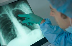
Methods of radiation diagnosis
In Phthisiology used X-ray and ultra sound techniques, radionuclide scanning, magnetic resonance imaging.In the differential diagnosis may be of value and positron emission tomography raphy (PET).
X-ray methods.For population screening and diagnosis of primary pulmonary and mediastinal chest x-rays are widely used. Other names of this method – fotorentgenografiya. as an image with X-ray pictures of the screen on film (plenoch Nye CT).Format standard modern pa kad 100 x 100 mm.
Compared with conventional X-ray fluorography can significantly increase the capacity of X-ray machine, cut spending on film and processing, to facilitate storage of the archive. Resolution method NOSTA fluorogram light quality is almost the same as the x-ray, so in some cases the fluorogram with an aspect ratio 100 x 100 mm on Zorn replaces x-ray of the lungs.Among the negative aspects of film fluorography home is the high radiation exposure to patients and staff.
Sienna on a film now comes to digital (digital) rentgenoflyuorografiya, which has many significant advantages. Chief among them – a high quality, and Jn formative possibility of computer processing iso map.Radiation exposure to the test in digital fluorography is 10-15 times lower than that of film (in the direct My projection respectively 0.05 and 0.7 mSv).It is also necessary to note the great speed of image acquisition, the combined view and print the paper a few images, transfer them to the distance, convenience store and then retrieve all the data, the low cost of the study.
Currently, digital rentgenoflyuorografiya gaining for follow-up surveys of large populations in order to timely diagnosed of tuberculosis, cancer and other diseases of the chest.She also successfully replaces X-ray survey went FIR as a diagnostic method.Russian industry produces laziness different models of digital scanning and pulse devices.
X-rays of light begin to review the image on the front line of the projection (with film magazine in front of piles of Noah wall).Pathological changes in the rear separated crystals of light to produce useful overview shot in the back of the line of projection (with cassette tape at the back of the chest wall).Then make a survey in the lateral projection images tion – the right and left.At the right side image to the box office those with adherent film of the right side of the chest at the left – the left.Radiographs in lateral projection tsiyah needed to determine the localization of the pathological process in the lung lobes and segments, identifying change equations in the interlobular clefts in the lungs and the shadows of the heart and diaphragm.When bilateral pulmonary disease better de lat in oblique projection images, which are obtained separate images of the right and left lung.
X-ray images are usually made at the height of inspiration. In the expiratory images make for better detection of edges and light kollabirovannogo pleural adhesions in the presence of pneumothorax, as well as to determine the displacement of mediastinal pathology of the lungs and pleura.
Increase the information content of X-ray exposure can change Niemi or rigidity of X-rays.Such images are called supereksponirovanpymi and rigid. They make patients with exudative pleurisy and pleural mi massive overlays, after surgery on the lungs, to better identify the walls of the trachea and bronze Hove.On supereksponirovannyh and hard shots can you manifest in areas of intense darkening of the various structures that were not visible on a conventional photograph.However, the intensity of the shadow of a low intensity in these images are not displayed.
Survey radiographs and lateral projections, if necessary, supplement the sighting shots a narrow beam of light. To do this, under the supervision of the patient rentgenotelevideniya give a position which enables it to bodit investigational lung field from interfering bone and other structures.
It should be noted that the radiological signs of some diseases are often so relief that the diagnosis only one experienced view of the radiograph.
X-rays produced, usually by using an image-enhancing X-ray images and zheniya rentgenotelevideniya.Used this method after X-rays for certain indications. Such explosion lyayutsya control during sighting shots and diagnostic České punctures, rentgenobronhologicheskih, angiographic and fistulograficheskih research.Fluoroscopy is needed to detect free fluid, moving in the pleural cavity, the determination of the mobility diameter and the fragmentation of the state of pleural sinuses.In many cases, fluoroscopic X-ray control better staring first days after intrathoracic surgery.However Finally, use fluoroscopy to evaluate the mobility of the diaphragm and of the samples with higher and lower pressure inside the thoracic (Valsalva maneuver, and Mueller, a symptom Golyzhnehta-Jacobson).Documentation of the results of these tests can be made and video footage rentgenokinosemkoy.
Computed tomography (CT) – Method rentgenologiche Skogen study, which has been universally recognized and applied in all fields of clinical medicine.CT provides images of cross sections brow vecheskogo body (axial view).X-ray tube rotates around the longitudinal axis of the patient’s body. Thin ny sheaf of rays from different angles passes through the investigated layer and captured many scintillation detectors, which move together with the tube.Different density tissues they pass through X-rays Chi, causes various changes in the intensity of the beam.It accurately recorded by detectors, processed by a computer and transformed on the image of the investigated cross-layer on a television eq wound.Thus, CT is no picture in the usual sense of the word, figure, made by com puter-based mathematical analysis of the degree of X-ray absorption tissues of different densities (computer tomography).
Conventional computed tomography scanning technology suggests a step by step movement of the table with the patient and how to stop x-ray tube after each cycle of rotation.They allow us to investigate cross-layer thickness schinoy from 2 to 10 mm.Scanning a single layer continues Xia few seconds.Significant increase in contrast can be obtained with intravenous contrast solution. Axial (transverse) images can be reconstructed using a computer to direct, lateral and oblique tomograms investigated area. Ravine bone and contrast can be changed within wide limits.CT Respiratory perform 6.12 standard slices. All results are parallel to the image on a television screen Niemi stored in computer memory and can be reproduced as an image on photographic paper or a Polaroid x-ray film.
A significant opportunity is the quantitative CT density of the tissue An estimate and media in the conventional unit in units of the scale Hounsfilda.The density of water on this scale is 0, air (-) 1000 units., Lightweight (+) 600 units.; Bone (+) 1000ed ..
In recent years the accepted methods to improve imaging in the study of light began to spiral CT and multiplanar. Helical CT technology is in constant rotation while the X-ray of the tube with a longitudinal movement of the patient.In this connection, instead of images of individual sections of the data collected from the total study area. During the full rotation of X ray tube depending on the pitch can be done by a different number of slices.
The advantages of the above methods of scanning – a significant reduction in time (from 10 to 20) and opportunity to study at a breath-hold.On exceeds the resolving power improves the quality of images of moving organs, creating favorable conditions for the study of children and seriously ill patients.Spiral Nye opened the way CT reconstruction and the creation of three-dimensional images of high quality.You can get a picture similar to bronchoscopic (computer bronhosko anisotropy), bronhograficheskimi (CT bronchography), while intravenous contrast – and angiographic (CT angiography).Reduced radiation exposure, since at least there is a need to clarify the repeated sections of diagnostic questions. With multiplanar imaging by increasing the number of detectors permit further improved by reducing the time of scanning, reducing artifacts and empowerment of processing the image.In general, the improved ray imaging techniques at different intrathoracic disease allow us to obtain three-dimensional image and more accurately assess the situation analysis tomicheskuyu, including availability, location and extent of pathological changes in the dynamics.CT can also provide high accuracy transtora Kalnoy biopsy and pleural puncture difficult.
Through the CT with a special image processing can be a virtual bronchoscopic view (Fig. 6.8.
Magnetic resonance imaging (MRI), many advantages of MRI are the basis for its use in the study of brain and spinal cord, bones and joints, large vessels thoracic cavity Noah, heart and other internal organs.One of the important advantages of the method is the lack of radiation exposure to patients and staff copper Qing.
The patient is placed on a desk scanner. Study area of the body is placed in a strong magnetic field.
It expands the protons in their direction and creates a magnetic moment in the tissues, oriented parallel to the external magnetic on any.When exposed to pulses that are sent first to the magnetic field perpendicular from the radio transmitting ka carcass, the total magnetic vector changes direction and begins to revolve around a new axis.The result is the induction of electric current in a receiving coil – the appearance of tion of magnetic resonance signal.He transforms of a special analyzer and transferred to the screen in black and white monitor.
Character images with MRI is mainly determined by the so-called relaxation time, proton density and tasks of the researcher. Here, by the relaxation of the T-1 takes the time during which recovers the first initial orientation of the protons, respectively, the external magnetic field.Relaxation of the T-2 – this time of weakening of the field created by radio-frequency pulse. Change nenie time between the rf pulses allows us to obtain images of different contrast and well-differentiated variety of tissues.There are also semi chenie images in different planes and the implementation of three-dimensional reconstruction.
Interpretation of the images in MRI is more difficult than usual for an absolute majority of physicians whose X-ray pictures.For example, air, bone, fibrous tissue has long time T-1, short T-2 and submitted to darken the image.
MRI is contraindicated if the patient kardiosti mulyatora or other metallic implant.Tion studies can be quite long, so hard to implement and critically ill children.
Angiopulmonografiya is staining and X-ray study of pulmonary artery and its wet wei.There are two main methods angiopulmonogra phy – the general and selective.
With a total angiopulmonografii contrast solution is introduced through the catheter into a vein hand, the superior vena cava or right heart chamber. X-rays of the product lead to serially special angiographic unit.This method requires a significant amount of contrast medium (50-60 ml), and usually does not provide a clear map iso pulmonary blood vessels, especially in pathological changes of the lungs.Amputation of vessels do not always reflect their true state.
Selective angiopulmonografiya technically more complicated, but is used more often. It is carried out after the category terizatsii corresponding branch pulmonary artery.Serial pictures were taken after the administration of 10-12 ml of solution con trastnogo matter.Typically, selective angiopulmonografiyu combined with registration of pressure in the pulmonary blood circulation and blood gas study.
Indications angiopulmonografii limited. Appl nyayut for the diagnosis of thrombosis and pulmonary embolism, as well as to determine the ability to continuously unfolding kollabirovannogo lung – as judged on the degree of vascular fibrosis.
Technical capabilities allow you to perform common angiopulmonografiyu in digital form with the introduction of a small amount into a vein contrast solution. At the same time computer processing allows high-quality video images.
Bronchial arteriagrafiya is catheterization, contrast radiography and bronchial arteries and their branches. The study was conducted under local anesthesia and control rentgenotelevideniya. A special needle to puncture mandates Rehn femoral artery below the inguinal crease. Mandre replace metallic conductor in which an artery in the injected radiopaque catheter with isolated curved tip.Then the conductor removed, and the conduction catheter ried out at the aorta.The tip of the catheter consistently seek out the mouth of the bronchial arteries and injected them into the catheter, and then dye (hypaque, Urografin, urotrast or their equivalents) at 35 ml / s in the amount of 5.12 ml. Productivity drive serial radiographs.
The main indication for bronchial arteriography is pulmonary hemorrhage of unknown etiology and localization tion.In such cases, the arteriogram may be identified and the expansion of abnormal tortuosity of the bronchial arte ry, out of contrast medium beyond them (ekstravazaiiya), focal or diffuse gipervaskulyarizatsiya, aneurysms bronchial arteries and their thrombosis, retrograde filling of peripheral branches of the pulmonary artery via arterio-arterial anastomoses.
Contraindications to the study: marked atherosclerosis, obesity patient, pulmonary heart disease.
Complication of bronchial arteriography may be re mat in the femoral artery puncture.A rare but cha zhelym complication is vascular lesion with spinal cord dysfunction of the lower limbs and organs tazo O.Prevention of complications is provided strictly subject to SHM methodological and technical principles and details of the study.
Bronchography. Contrast X-ray studies of bronchial tion carried out under local anesthesia in the form of positional (non-directional) or selective (directed Noah) bronchography.In positional bronchography catheter is introduced into the trachea through the nose, during the administration of contrast material the patient’s body give the optimum position. Se lective catheterization based on bronchography investigated ICDO bronchus.To use a variety of designs for catheters and techniques.
Previously, bronchography was used very widely. At the present time due to the widespread use of CT, this method has lost its former significance.
Plevrografiya allows contrast and clarify the verge of particle purulent cavities in patients with pleural empyema.Initially produce a pleural puncture and aspirate the pleural contents. Then, under the control of rentgenotelevideniya injected into the pleural cavity of 30-40 ml of warm X-ray solution (propyliodone, Urografin). Rentgenogram we are doing in different projections, changing the position of the patient.After completing studies with contrast material balances kami pleural contents aspirated.Information, which is reached at plevrografii, in most instances s can be obtained using CT.
Fistulografiyu used for examination of patients with various thoracic and torakobronhialnymi fistula. Before you install the appropriate sensing fistulografiey Niemi direction of the fistulous.Contrast agent injected into the fistulous course syringe through the catheter under the control of rentgenotelevideniya. Apply oil or vodorastvo rimye radiopaque agents.Then produce X-rays in different projections, changing the position of Nogo pain, or a CT scan, in-process research and after analyzing the images reveal the anatomical features of the fistula, determine its message with the pleural cavity and bronchi Alno tree.In the case of penetration of contrast agent into the bronchial tree is obtained
retrograde ISF tulobroihografiya.After completing the study pre Paraty through a catheter to suction capabilities, and offer the patient a good cough.
Ultrasound techniques, particularly ultrasound, different security, the possibility of multiple studies, high resolution.
TB in the practice of ultrasonic methods are useful for accurate determination and control over the size of peripheral lymph nodes (cervical, podmyshech GOVERNMENTAL, groin).With the help of ultrasound can determine the difference in fluid in the pleural cavity, since its presence ence between the parietal pleura and slight hypo-echogenic marked area.Ultrasonic testing is to select a point for the puncture of the pleural cavity. After pneumonectomy dynamic detection of fluid in the pleural cavity often can replace X-ray studies tion.
An important and often crucial ultrasound is the examination of men and women with suspicious rhenium on tuberculosis of the urogenital tract.It is not required as to monitor the dynamics of the process for the treatment and ftiziouroloptcheskih ftizioginekologicheskih patients.
Radionuclide (radioisotope) methods have a leading role for regional assessment of ventilation and blood flow in the lungs. They are based on inhalation or bowl inside a Venn introduction radiopharmaceuticals labeled GOVERNMENTAL gamma-emitting radionuclides.This xenon-excited sultry blend of (133 Xe), macroaggregates of albumin (I31 I or 99m Tc), indium citrate (133m In), albumin microspheres (99m Tc or 133m In), etc.Registration of the drug distribution is carried out with the help of a scintillation gamma camera with a computer.At the same time can be both static and dynamic scintigraphy in the front, rear and side O projections.All parameters are usually determined as a percentage max, respectively, dividing the lung fields on the upper, middle and lower zones.However, mathematical modeling tion allows to evaluate the ventilation and blood flow in the lungs and in absolute terms.
The study of regional lung function radionuclide methods should be conducted prior to X-ray studies Nij.The information obtained can be judged not only on ventilation and blood flow, but also on the localization and propagation of gravity changes in the lungs.
New perspectives in the studies of pulmonary ventilation and circulation, open MRI. Currently, this method is beginning to be used to assess ventilation various parts of the lungs after inhalation of hyperpolarized helium prior.
Pozitropnaya emission tomography (PET) is spreading in the differential diagnosis of intrapulmonary structures, based on the assessment of cellular PET ME tabolizma.Intravenous drug radiofarmakologichesky FDG (18 F-flyuorodeoksiglyukoza), which is sensitive to increased metabolism of glucose in cancer cells in semiconductors and scans forms a bright spot.Cancer cells can be detected in a lymph node diameter of less than 1 cm rum.Informativeness of PET increases with its combination with CT and creating a composite image.


